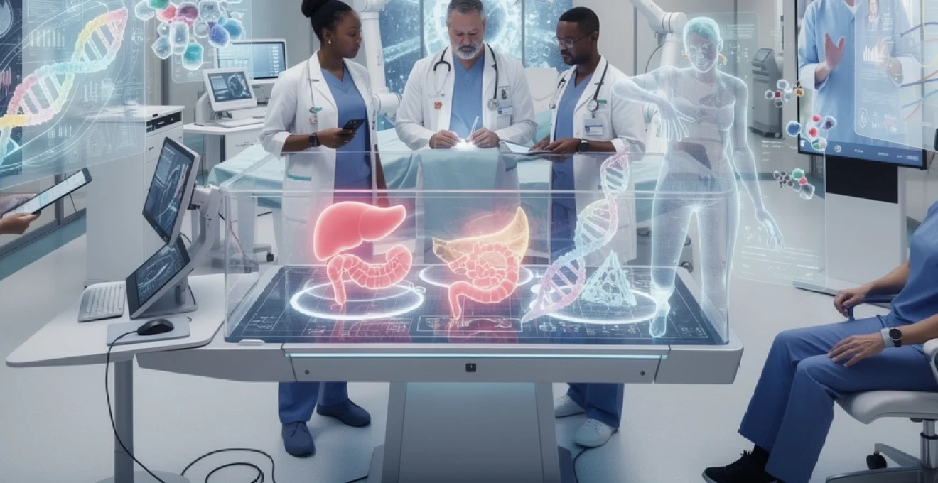
# Exploring Technological Innovations in Medicine and Patient Care
The landscape of modern healthcare has undergone a remarkable transformation over the past decade, driven by exponential advances in computational power, materials science, and molecular biology. From operating theatres equipped with robotic surgeons to smartphone applications that monitor cardiac rhythms in real-time, technology has fundamentally reshaped how medical professionals diagnose, treat, and manage disease. These innovations extend far beyond simple digitisation—they represent a paradigm shift towards precision medicine, where treatments are tailored to individual genetic profiles, and preventive care is guided by predictive algorithms that identify risk factors years before symptoms emerge. The convergence of artificial intelligence, nanotechnology, and genomic editing has created unprecedented opportunities to address conditions once deemed incurable, whilst simultaneously improving accessibility to quality healthcare for populations in remote or underserved regions.
Artificial intelligence and machine learning in clinical diagnostics
The integration of artificial intelligence into clinical diagnostics has fundamentally altered the accuracy and speed with which medical professionals can identify disease. Machine learning algorithms, trained on vast datasets comprising millions of patient records and medical images, now demonstrate diagnostic capabilities that match or exceed human expert performance across multiple specialties. These systems don’t simply replicate human decision-making—they identify patterns invisible to the human eye, processing information at scales and speeds that would be impossible for even the most experienced clinicians. The economic implications are equally significant, with AI-driven diagnostics potentially reducing healthcare costs by billions annually through earlier disease detection and more efficient resource allocation.
Deep learning algorithms for radiology image analysis and tumour detection
Convolutional neural networks have revolutionised radiological interpretation, particularly in oncology where early tumour detection dramatically improves survival rates. These deep learning systems analyse CT scans, MRIs, and mammograms with remarkable precision, identifying subtle abnormalities that human radiologists might overlook. Recent studies demonstrate that AI algorithms can detect breast cancer from mammograms with 99% accuracy whilst reducing false positives by up to 30%. The technology excels at identifying microcalcifications and architectural distortions—early indicators of malignancy that often escape initial visual inspection. Beyond simple detection, these algorithms can classify tumour types, predict growth rates, and even suggest optimal biopsy locations, transforming radiology from a purely diagnostic specialty into a predictive and therapeutic planning discipline.
The efficiency gains are equally compelling. A single AI system can analyse thousands of scans in the time it takes a radiologist to review dozens, dramatically reducing waiting times for patients anxiously awaiting results. This capability proves particularly valuable in resource-constrained settings where radiologist shortages create dangerous backlogs. Furthermore, these systems continuously learn and improve, incorporating new data from every scan they analyse to refine their diagnostic accuracy. The technology also provides consistency that human interpretation cannot guarantee—fatigue, distraction, and cognitive biases don’t affect algorithmic analysis, ensuring every patient receives the same standard of care regardless of when their scan is reviewed.
IBM watson health and natural language processing for electronic health records
Natural language processing technologies have transformed how healthcare systems extract actionable insights from the vast repositories of unstructured data contained within electronic health records. IBM Watson Health exemplifies this capability, employing sophisticated algorithms to parse physician notes, laboratory reports, and patient histories to identify critical information that might otherwise remain buried in text. The system can rapidly review thousands of pages of medical literature alongside a patient’s complete medical history, suggesting evidence-based treatment options that align with the latest research findings. This capacity for comprehensive data synthesis enables clinicians to make more informed decisions, particularly when treating complex conditions requiring multidisciplinary approaches.
The technology addresses one of healthcare’s most persistent challenges: information overload. With medical knowledge doubling approximately every 73 days, remaining current across all relevant research becomes humanly impossible. Watson Health bridges this gap by continuously monitoring new publications, clinical trials, and treatment outcomes, then contextualising this information within individual patient scenarios. The system also identifies potential drug interactions, contraindications based on genetic markers, and alternative therapeutic approaches that might not immediately occur to treating physicians. This augmentation of clinical decision-making doesn’t replace medical expertise—it amplifies it, providing physicians with computational support that enhances rather than supplants their professional judgement.
Predictive analytics in sepsis detection and early warning systems
Sepsis represents one of medicine’s most urgent challenges, with mortality rates increasing significantly for every hour
of delayed recognition. Traditional early warning scores rely on periodic vital sign checks and manual chart reviews, which can miss subtle but dangerous trends. Predictive analytics platforms continuously ingest data from bedside monitors, laboratory systems, and electronic health records to identify complex patterns that precede clinical deterioration. By analysing variables such as heart rate variability, lactate levels, respiratory rate, and changes in mental status in real time, these AI-driven sepsis detection tools can alert clinicians hours before overt organ failure develops.
Hospitals deploying machine learning–based early warning systems report reductions in sepsis-related mortality of up to 20–30%, alongside shorter intensive care unit stays. Importantly, the most effective solutions are tightly integrated into existing clinical workflows, presenting risk scores and alerts within the EHR rather than as separate dashboards that clinicians must remember to check. To avoid “alarm fatigue”, advanced models calibrate their thresholds dynamically, adjusting to local patient populations and incorporating clinician feedback to improve precision over time. As these systems mature, we are likely to see predictive analytics extend beyond sepsis to encompass broader deterioration syndromes, creating a safety net that continuously watches over patients on the ward.
Google DeepMind’s AlphaFold for protein structure prediction in drug discovery
While many AI applications in healthcare focus on clinical data, Google DeepMind’s AlphaFold has transformed an entirely different domain: structural biology. For decades, predicting a protein’s three-dimensional structure from its amino acid sequence was a grand challenge in science, often requiring years of painstaking experimental work using X-ray crystallography or cryo-electron microscopy. AlphaFold’s deep learning models can now predict protein structures with near–experimental accuracy in a matter of hours, compressing years of lab effort into a single computational run. This breakthrough has profound implications for drug discovery, where understanding protein shape is essential for designing molecules that can bind to disease targets with high specificity.
Pharmaceutical companies and academic laboratories are already leveraging AlphaFold’s freely available structural predictions to accelerate target validation and hit discovery. For example, by knowing the precise configuration of a viral enzyme or an oncogenic receptor, chemists can use structure-based drug design to identify candidate compounds that fit like keys into highly specific molecular locks. This reduces the number of blind screening experiments required, saving both time and cost. Just as digital maps revolutionised navigation, AlphaFold’s protein structure atlas is rapidly becoming a foundational reference for modern biomedicine, guiding researchers through the complex terrain of cellular machinery and enabling more rational, personalised therapies.
Robotic-assisted surgery and minimally invasive procedures
Robotic-assisted surgery represents one of the most visible intersections between engineering and medicine, transforming the way complex operations are performed. Rather than replacing surgeons, these systems act as precision-enhancing tools, filtering out tremor and scaling down hand movements to millimetre-level accuracy. Patients benefit from smaller incisions, reduced blood loss, and shorter hospital stays compared with traditional open procedures, while surgeons gain enhanced visualisation and ergonomic advantages that can extend their careers. As computing power and imaging technologies continue to improve, robotic platforms are evolving from mechanical extensions of the surgeon’s hands into intelligent collaborators capable of assisting with decision-making in real time.
Da vinci surgical system for precision laparoscopic operations
The da Vinci Surgical System is the most widely adopted robotic platform for minimally invasive surgery, used across specialties including urology, gynaecology, colorectal surgery, and thoracic oncology. Operating through a few small ports rather than a single large incision, da Vinci’s articulated instruments provide seven degrees of freedom, surpassing the dexterity of the human wrist inside the confined spaces of the body. Surgeons control these instruments from a console featuring a high-definition, three-dimensional view of the operative field, magnified up to tenfold. This enhanced visualisation allows for meticulous dissection around critical structures such as nerves and blood vessels, reducing the risk of complications.
Clinical studies have demonstrated that robotic prostatectomy with da Vinci is associated with lower blood loss, fewer transfusions, and quicker return of continence and sexual function compared with conventional approaches. Yet the system’s benefits are not purely clinical: ergonomically, it allows surgeons to operate seated, with reduced strain on the neck and shoulders, potentially mitigating the musculoskeletal injuries that plague many high-volume operators. For hospitals considering adoption, key success factors include structured training programmes, careful case selection during the learning curve, and close monitoring of outcome metrics to ensure that the promised gains in precision laparoscopic operations translate into real-world patient benefit.
Haptic feedback technology and surgeon-console interface design
One of the early limitations of robotic surgery was the loss of tactile sensation—surgeons could see tissue but not feel its resistance, which is crucial when judging how much force to apply. New generations of robotic systems are addressing this through advanced haptic feedback technology, which simulates touch by translating instrument-tip forces into subtle vibrations or resistance at the surgeon’s controls. This tactile information, combined with visual cues, helps operators distinguish between healthy tissue, scar tissue, and critical structures such as arteries. A well-designed surgeon-console interface becomes akin to a musical instrument: intuitive enough to disappear from conscious thought, allowing the surgeon to “play” complex procedures with focus and finesse.
Console design goes beyond haptics to encompass hand-controller geometry, foot-pedal layout, and the arrangement of on-screen information. Poorly designed interfaces can increase cognitive load, slow reaction times, and contribute to errors. By contrast, interfaces based on human factors research streamline task flow, surface the most relevant data at the right moment, and provide clear visual alerts when system parameters drift outside safe ranges. As you evaluate robotic solutions—whether as a clinician or hospital administrator—paying attention to interface usability and haptic feedback quality can be just as important as headline specifications like instrument range of motion or camera resolution.
Autonomous surgical robots and computer vision integration
While today’s surgical robots are largely teleoperated, research is rapidly progressing towards partial autonomy, where robots can complete defined subtasks under human supervision. Using computer vision and real-time image analysis, autonomous systems can recognise anatomical landmarks, track tissue deformation, and maintain instrument positioning with sub-millimetre accuracy. Early demonstrations have shown robots autonomously suturing soft tissues or maintaining a steady endoscopic view during dynamic procedures, freeing the surgeon to focus on higher-level decision-making. Think of this as similar to advanced driver-assistance systems in cars: the human remains in control, but software handles repetitive or highly precise manoeuvres.
Computer vision algorithms enable these robots to interpret surgical scenes much like self-driving cars interpret roads, segmenting organs, vessels, and instruments frame by frame. As datasets of labelled surgical videos grow, models will become more adept at predicting the next steps in a procedure and offering context-aware suggestions—such as recommending optimal incision sites or warning when instrument trajectories approach critical structures. Ethical and regulatory frameworks will play a decisive role in determining how quickly we move from assistive autonomy to more independent robotic actions, but the trajectory is clear: surgery is becoming a human–machine collaboration where each partner contributes their strengths.
Microsurgical robotics for ophthalmic and neurosurgical applications
At the frontiers of precision medicine, microsurgical robots are enabling interventions that push beyond the limits of human steadiness. In ophthalmology, robotic platforms have been used experimentally to inject therapeutic agents into the subretinal space and to manipulate tiny retinal vessels with a precision measured in microns. Such accuracy is essential when treating conditions like macular degeneration or diabetic retinopathy, where millimetre-scale errors can mean the difference between preserved and lost vision. Similarly, in neurosurgery, robot-assisted systems guide instruments through narrow, delicate corridors in the brain, minimising damage to surrounding tissue while accessing deep-seated tumours or aneurysms.
These microsurgical systems often incorporate advanced imaging modalities—such as intraoperative OCT (optical coherence tomography) or high-resolution MRI guidance—to provide real-time feedback on instrument position relative to critical structures. For patients, the promise is fewer neurological deficits and faster recovery; for surgeons, the benefit lies in a level of precision and stability that simply cannot be matched by unaided hands. As these technologies mature, we may see them combined with augmented reality overlays and AI-based navigation to create “GPS for surgery”, guiding microscale manoeuvres in some of the body’s most sensitive territories.
Telemedicine platforms and remote patient monitoring technologies
Telemedicine has moved from a niche service to a central pillar of modern healthcare delivery, accelerated dramatically by the COVID-19 pandemic. By decoupling clinical expertise from physical location, virtual care platforms make it possible for patients to consult specialists without leaving their homes and for clinicians to monitor chronic diseases between clinic visits. This shift is not just about convenience; it’s about reimagining care as a continuous, data-driven relationship rather than a series of episodic encounters. When combined with remote patient monitoring technologies, telemedicine enables earlier intervention, reduces hospital admissions, and enhances patient engagement in their own health.
Real-time vital signs tracking through IoMT-enabled wearable devices
The Internet of Medical Things (IoMT) connects wearable devices, home sensors, and medical-grade monitors into an integrated network that streams health data in real time. Smartwatches and patches now routinely track heart rate, oxygen saturation, sleep patterns, and physical activity, while more specialised devices can monitor arrhythmias, blood pressure, or glucose levels continuously. For patients with conditions such as heart failure, COPD, or diabetes, this always-on monitoring provides a safety net: deviations from baseline can trigger automated alerts to clinical teams, prompting timely adjustments to medication or lifestyle before a crisis develops.
From a practical standpoint, the most successful remote monitoring programmes focus on actionable data rather than simply collecting numbers. Dashboards that highlight trends, out-of-range values, and adherence patterns help clinicians quickly identify who needs attention. For patients, intuitive mobile apps that translate metrics into plain-language insights—such as “your average nightly sleep has dropped by 90 minutes this week”—make it easier to understand and act on recommendations. As you consider adopting IoMT-enabled wearable devices, key questions include data accuracy, battery life, ease of use, and how seamlessly the data integrate into existing telemedicine platforms and electronic health records.
5G network infrastructure for high-resolution video consultations
High-quality telemedicine relies on robust connectivity. The rollout of 5G network infrastructure, with its high bandwidth and low latency, is unlocking new possibilities for remote care that simply were not feasible over earlier networks. With 5G, clinicians can conduct high-resolution video consultations where subtle visual cues—skin colour changes, fine motor tremors, facial asymmetries—are reliably visible, improving diagnostic accuracy. In more advanced scenarios, 5G can support the real-time streaming of medical imaging, enabling remote specialists to guide procedures or interpret scans as they are acquired.
Low-latency connections are particularly important for emerging applications such as remote robotic procedures or synchronous tele-ultrasound, where a clinician in one location manipulates a probe or instrument in another. While these scenarios are still relatively rare, they illustrate how connectivity is becoming a clinical safety issue rather than a purely technical concern. For health systems, investing in secure, resilient network infrastructure—and ensuring equitable access in rural or underserved areas—will be critical to realising the full potential of telemedicine and high-resolution video consultations.
Cloud-based electronic health record interoperability standards
One of the persistent barriers to effective telemedicine is fragmentation of patient data across multiple, incompatible systems. Cloud-based electronic health record (EHR) platforms, built around interoperability standards such as FHIR (Fast Healthcare Interoperability Resources), aim to solve this by enabling secure, standards-based exchange of information between providers, devices, and applications. When an EHR can seamlessly pull in teleconsultation notes, remote monitoring data, imaging reports, and lab results, clinicians gain a holistic view of the patient’s health regardless of where care took place.
Interoperability also empowers patients, who increasingly expect to access and share their health information as easily as they manage their banking or travel bookings. Cloud-based architectures make it possible to build patient-facing portals and mobile apps that connect to a unified record, supporting tasks such as appointment scheduling, prescription refills, and secure messaging. Of course, this connectivity raises important questions around data privacy and cybersecurity. Implementing strong encryption, fine-grained access controls, and rigorous audit trails is essential to ensure that the benefits of cloud-based EHR interoperability do not come at the expense of patient trust.
Remote diagnostics using store-and-forward asynchronous teledermatology
Not all telemedicine needs to happen in real time. Store-and-forward models, where clinical data are captured and reviewed later, can be highly efficient for specialties such as dermatology. In asynchronous teledermatology, patients or primary care providers upload high-resolution images of skin lesions along with brief clinical histories to secure platforms. Dermatologists then review these cases in batches, providing diagnoses and management recommendations without the need for a live video connection. This approach dramatically reduces waiting times, particularly in areas where specialist access is limited.
To ensure diagnostic quality, best practice includes standardised image capture guidelines—covering lighting, focus, and lesion framing—as well as structured data fields for symptoms, duration, and risk factors. Some platforms now integrate AI-based pre-screening tools that flag lesions with features suggestive of malignancy, helping clinicians prioritise urgent cases. For health systems, asynchronous teledermatology offers a practical way to expand specialist reach, optimise dermatologist time, and reduce unnecessary in-person referrals, while still ensuring that patients with suspicious or complex lesions are seen face-to-face when necessary.
CRISPR gene editing and personalised genomic medicine
CRISPR-Cas9 gene editing has moved from theoretical promise to clinical reality in just over a decade, offering unprecedented control over the human genome. At its core, CRISPR acts like a molecular scalpel, guided by a short piece of RNA to cut DNA at a specific sequence. Once the break is made, cellular repair mechanisms can be harnessed to disable faulty genes, correct point mutations, or insert therapeutic sequences. For monogenic disorders such as sickle cell disease or certain forms of inherited blindness, this means we can now contemplate one-time treatments that address the root genetic cause rather than merely managing symptoms.
Personalised genomic medicine builds on this capability by tailoring interventions to each individual’s unique DNA profile. Whole-genome sequencing can reveal predispositions to drug toxicity, disease risk, or differential treatment response, allowing clinicians to select therapies with a higher likelihood of success and fewer adverse effects. In oncology, for example, tumour sequencing informs the use of targeted therapies and immunotherapies that exploit specific molecular vulnerabilities. As CRISPR-based therapies progress through clinical trials, integrating genomic data into routine care will require robust bioinformatics pipelines, clear consent processes, and careful consideration of the ethical implications of editing the human germline versus somatic cells.
Augmented reality and virtual reality in medical training and surgical planning
Augmented reality (AR) and virtual reality (VR) technologies are redefining how medical professionals learn, rehearse, and execute complex procedures. VR immerses trainees in fully simulated environments where they can practise everything from basic laparoscopic skills to managing rare emergency scenarios, all without risk to real patients. AR, by contrast, overlays digital information onto the real world, allowing surgeons to see three-dimensional reconstructions of a patient’s anatomy superimposed on their body during preoperative planning or even in the operating room. Together, these tools function like advanced flight simulators for medicine, enabling experiential learning that is more engaging and effective than traditional textbooks or lectures.
Studies consistently show that VR-based training improves procedural accuracy, reduces operative time, and boosts learner confidence compared with conventional methods. For surgical planning, AR can project CT or MRI-derived models onto the patient, helping teams visualise tumour margins, vascular anatomy, or spinal alignment before making the first incision. This leads to more precise interventions and may reduce the need for intraoperative imaging. As hardware becomes more affordable and software platforms more user-friendly, we can expect AR and VR to become standard components of curricula in medical schools and residency programmes, levelling the training playing field between large academic centres and smaller hospitals.
Nanotechnology applications in drug delivery and regenerative therapies
Nanotechnology operates at a scale where materials exhibit unique physical and chemical properties, opening new avenues for treating disease. In drug delivery, nanoparticles can be engineered to carry therapeutic agents directly to diseased tissues while sparing healthy cells, much like targeted delivery drones. By adjusting size, surface charge, and coating molecules, researchers can design nanocarriers that evade immune detection, cross biological barriers such as the blood–brain barrier, and release their cargo in response to specific triggers like pH changes or enzyme activity. This level of precision holds particular promise for oncology, where traditional chemotherapy often causes significant collateral damage.
Beyond drug delivery, nanomaterials are playing a growing role in regenerative medicine. Scaffolds constructed from biocompatible nanofibres can mimic the extracellular matrix, providing a supportive framework for stem cells to grow and differentiate into functional tissues. For example, nanostructured surfaces on orthopaedic implants encourage bone cells to integrate more tightly, improving long-term stability. In cardiology, injectable hydrogels containing nanoparticles and growth factors are being investigated to promote tissue repair after myocardial infarction. As with any powerful technology, careful long-term safety studies are essential to understand how nanomaterials interact with the body over time, but early results suggest that nanotechnology will be a cornerstone of future therapies aimed at not just treating disease, but rebuilding damaged organs and tissues.